Lower Nose Arterial Plexus and Implications for Safe Filler Injections (2022)
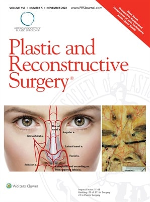
Title : Lower Nose Arterial Plexus and Implications for Safe Filler Injections
Researcher : Tansatit, T., Jitaree, B., Uruwan, S., Rungsawang, C.
Abstract : Background: The lower nose has abundant blood supply; however, nasal tip necrosis still occurs following filler injections. This study revealed the complicated pattern of the arterial supply of the lower nose.
Methods: The arterial pattern of the lower nose was studied in 40 cadavers using conventional dissections and translucent modified Sihler staining.
Results: Two arterial rings were connected in a figure of eight. The upper ring (nasal arterial circle) consisted of the lateral nasal and the subalar arteries encircling the nasal tip and alae. The lower ring (arterial plexus of the upper lip) was more important because of the contribution of the facial and superior labial arteries. This specific feature had not been mentioned elsewhere.
Conclusion: Understanding this specific feature of the blood supply of the lower nose is essential for aesthetic physicians to perform the appropriate techniques during filler injection procedures in the nasal and perioral regions.
Link to Academic article: DOI: 10.1097/PRS.0000000000009649
Journal : Plastic and Reconstructive Surgery, 2022, 150(5),
Bibliography : Tansatit, T., Jitaree, B., Uruwan, S., & Rungsawang, C. (2022). Lower Nose Arterial Plexus and Implications for Safe Filler Injections. Plastic and Reconstructive Surgery,150(5), 987E–992E. DOI: 10.1097/PRS.0000000000009649

The analgesic efficacy of anterior femoral cutaneous nerve block in combination with femoral triangle block in total knee arthroplasty: a randomized controlled trial(2021)
Title : The analgesic efficacy of anterior femoral cutaneous nerve block in combination with femoral triangle block in total knee arthroplasty: a randomized controlled trial
Researcher : Kampitak, W., Tanavalee, A., Tansatit, T., …Songborassamee, N., Vichainarong, C.
Abstract : Background: Ultrasound-guided femoral triangle block (FTB) can provide motor-sparing anterior knee analgesia. However, it may not completely anesthetize the anterior femoral cutaneous nerve (AFCN). We hypothesized that an AFCN block (AFCNB) in combination with an FTB would decrease pain during movement in the immediate 12 h postoperative period compared with an FTB alone.
Methods: Eighty patients scheduled to undergo total knee arthroplasty were randomized to receive either FTB alone (FTB group) or AFCNB with FTB (AFCNB + FTB group) as part of the multimodal analgesic regimen. The primary outcome was pain during movement at 12 h postoperatively. Secondary outcomes included numeric rating scale (NRS) pain scores, incidence of surgical incision site pain, intravenous morphine consumption, immediate functional performance, patient satisfaction, and length of hospital stay.
Results: The NRS pain scores on movement 12 h postoperatively were significantly lower in the AFCNB + FTB group than in the FTB group (mean difference: –2.02, 95% CI: –3.14, –0.89, P < 0.001). The incidence of pain at the surgical incision site at 24 h postoperatively and morphine consumption within 48 h postoperatively were significantly lower (P < 0.001), and quadriceps muscle strength at 0° immediately after surgery was significantly greater in the AFCNB + FTB group (P = 0.04).
Conclusions: The addition of ultrasound-guided AFCNB to FTB provided more effective analgesia and decreased opioid requirement compared to FTB alone after total knee arthroplasty and may enhance immediate functional performance on the day of surgery.
Keywords: Arthroplasty; Knee; Nerve block; Peripheral nerves; Postoperative pain; Ultrasonography.
Link to Academic article: DOI: https://doi.org/10.4097/kja.21120
Journal : Korean Journal of Anesthesiology, 2021, 74(6).
Bibliography : Kampitak, W., Tanavalee, A., Tansatit, T., Ngarmukos, S., Songborassamee, N., & Vichainarong, C. (2021). The analgesic efficacy of anterior femoral cutaneous nerve block in combination with femoral triangle block in total knee arthroplasty: a randomized controlled trial. Korean Journal of Anesthesiology, 74(6), 496–505. DOI: https://doi.org/10.4097/kja.21120

The crest injection technique for glabellar line correction and the paracentral artery (2021)
Title : The crest injection technique for glabellar line correction and the paracentral artery
Researcher : Tansatit, T., Uruwan, S., Rungsawang, C.
Abstract : The glabella is a zone that carries a high risk of blindness after performing filler injections. The arteries beneath the glabellar lines were investigated by meticulous dissections in 30 geriatric embalmed cadavers with latex injections into the arterial system. The results showed that the supratrochlear artery, a direct branch of the ophthalmic artery, ascended from the muscular layer of the medial eyebrow along the medial canthal vertical line of the intercanthal vertical zone (53 in 60 hemifaces, or 88%). The dominant single paracentral artery from the radix artery was found within the radix vertical zone (eight out of 30 glabellae, or 27%). Among these, the dominant paracentral artery was near the midline in two cadavers and arose along the radix vertical line in six cadavers. The dominant paracentral artery may be the cause of ocular complications during injections of glabellar lines between the medial eyebrows, especially at the radix vertical lines. The supratrochlear artery might cause ocular complications when an injection is performed close to the medial eyebrows. Pinching to create a skin crest and evert glabellar line for a precise injection is recommended to temporarily occlude the paracentral artery.
Link to Academic article: DOI: 10.1097/GOX.0000000000003982
Journal : Plastic and Reconstructive Surgery – Global Open, 2021, 9(12).
Bibliography : Tansatit, T., Uruwan, S. & Rungsawang, C.,(2021). The crest injection technique for glabellar line correction and the paracentral artery. Plastic and Reconstructive Surgery – Global Open, 9(12), e3982. DOI: 10.1097/GOX.0000000000003982

The feasibility determination of risky severe complications of arterial vasculature regarding the filler injection sites at the tear trough (2018)
Title : The feasibility determination of risky severe complications of arterial vasculature regarding the filler injection sites at the tear trough
Researcher : Jitaree, B., Phumyoo, T., Uruwan, S., …McCormick, L., Tansatit, T.
Abstract : Background: The tear trough is a significant sign of periorbital aging and has usually been corrected with filler injection. However, the arterial supply surrounding the tear trough could be inadvertently injured during injection; therefore, this study aimed to evaluate the nearest arterial locations related to the tear trough and investigate the possibility of severe complications following filler injection.
Methods: Thirty hemifaces of 15 Thai embalmed cadavers were used in this study.
Results: The artery located closest to both the inferior margin (TT1) and mid-pupil level (TT2) of the tear trough was found to be the palpebral branch of the infraorbital artery. Furthermore, at 0.5 mm along the tear trough from the medial canthus (TT3), the angular artery was identified, which was found to be a branch of the ophthalmic artery. The artery at TT1 and TT2 was located beneath both the zygomaticus major and the orbicularis oculi muscles. The distances from TT1 to the artery were measured as follows: laterally, 2.79 ± 1.08 mm along the x axis; and inferiorly, 2.88 ± 1.57 mm along the y axis. For the TT2, the artery was located inferomedially from the landmark of 4.65 ± 1.83 mm along the x axis and 7.13 ± 3.99 mm along the y axis. However, the distance along the x axis at TT3 was located medially as 4.00 ± 2.37 mm.
Link to Academic article: DOI: 10.1097/PRS.0000000000004893
Journal : Plastic and Reconstructive Surgery, 2018, 142(5).
Bibliography : Jitaree, B., Phumyoo, T., Uruwan, S., Sawatwong, W., McCormick, L., & Tansatit, T. (2018). The feasibility determination of risky severe complications of arterial vasculature regarding the filler injection sites at the tear trough. Plastic and Reconstructive Surgery, 142(5), 1153–1163. DOI: 10.1097/PRS.0000000000004893