Nongenomic action of aldosterone on colocalization of angiotensin II type 1 and type 2 receptors in rat kidney (2018)
Title : Nongenomic action of aldosterone on colocalization of angiotensin II type 1 and type 2 receptors in rat kidney
Researcher : Sinphitukkul, K., Manotham, K., Eiam-Ong, S., Eiam-Ong, S.
Abstract : Previous in vitro studies have demonstrated that angiotensin II type 1 and type 2 receptors (AT1R and AT2R) are co-localized and can form AT1R/AT2R dimerization in rat proximal tubular cells. Aldosterone non-genomically enhances angiotensin II receptor dimerization. We found no other in vivo studies in the literature regarding the effect of aldosterone on colocalization of AT1R and AT2R in whole kidney. Male Wistar rats were intraperitoneally injected with either normal saline solution (sham group) or aldosterone (experimental group). Colocalization of renal AT1R and AT2R proteins was examined by double immunohistochemical staining. The colocalization of AT1R and AT2R proteins was more prominent in the glomerulus, distal convoluted tubules, and cortical collecting ducts while colocalization was weak and diffused in the proximal convoluted tubules and peritubular capillaries in both groups. Our in vivo study showed aldosterone did not alter a constitutive colocalization of AT1R and AT2R proteins in the renal cortex and medulla. However, these proteins were colocalized more prominently in the renal cortex.
Keywords: Aldosterone, angiotensin II receptors, nongenomic action, protein colocalization, rat kidney
Link to Academic article: https://doi.org/10.1080/01478885.2018.1438756
Journal : ,
Bibliography : Sinphitukkul, K., Manotham, K., Eiam-Ong, S., & Eiam-Ong, S. (2018). Nongenomic action of aldosterone on colocalization of angiotensin II type 1 and type 2 receptors in rat kidney.
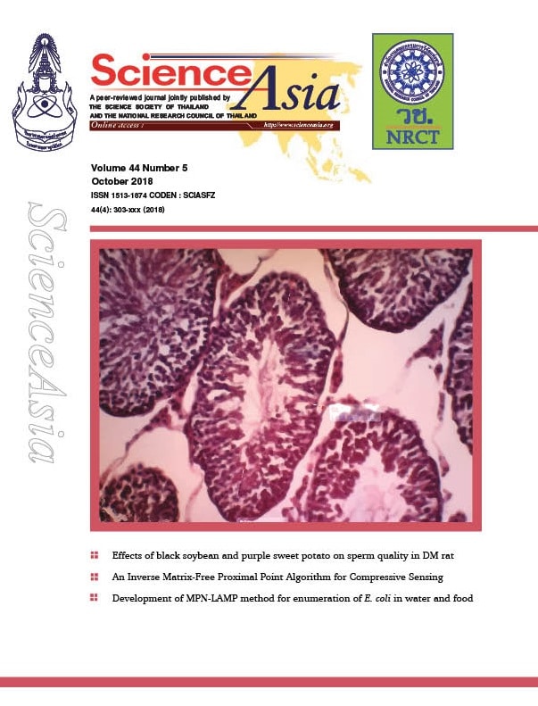
Nongenomic effect of aldosterone on angiotensin II type 1 receptor dimerization in human renal proximal tubular cells: Implications for endoplasmic reticulum stress (2018)
Title : Nongenomic effect of aldosterone on angiotensin II type 1 receptor dimerization in human renal proximal tubular cells: Implications for endoplasmic reticulum stress
Researcher : Sinphitukkul, K., Manotham, K., Eiam-Ong, S., …Inagi, R., Eiam-Ong, S.
Abstract : In vitro studies have showed that aldosterone increases oxidative stress molecules through a nongenomic effect. Oxidative stress induces angiotensin II type 1 receptor (AT1R) dimerization and endoplasmic reticulum (ER) stress, leading to renal tubular damage. However, the nongenomic effect of aldosterone on AT1R dimerization and ER stress in renal cells has not been determined. Here, we examined the nongenomic action of aldosterone in renal proximal tubular epithelial cells (PTECs) to better understand the underlying mechanisms. HK-2 cells, human renal PTECs, were exposed to vehicle or aldosterone for 30 min. In two additional groups, the cells were pretreated with eplerenone, a mineralocorticoid receptor (MR) blocker or apocynin, an NADPH oxidase inhibitor, for 30 min before aldosterone incubation. Protein abundances of dimeric/monomeric forms of AT1R, p47phox (a cytosolic part of NADPH oxidase), and activating transcription factor 4 (ATF4), a transcription factor responsive to ER stress, were determined by Western blotting. Aldosterone nongenomically increased plasma membrane protein expression of AT1R dimeric forms in a time- and dose-dependent manners. The levels of the cytosolic p47phox protein declined while the membranous protein level was enhanced following aldosterone treatment. The aldosterone induced alteration in these two proteins was abolished by pretreatment with eplerenone or apocynin. In addition, aldosterone (100 nM) induced nuclear ATF4 protein accumulation in a time-dependent fashion, which was blocked by apocynin and partially attenuated by eplerenone. Aldosterone nongenomically increased AT1R dimerization and nuclear ATF4 protein accumulation dependent on MR and NADPH oxidase activation. Hence aldosterone could induce AT1R dimerization and activate the endoplasmic reticulum stress response.
Link to Academic article: doi: 10.2306/scienceasia1513-1874.2018.44.332
Journal : ,
Bibliography : Sinphitukkul, K., Manotham, K., Eiam-Ong, S., Nangaku, M., Inagi, R. & Eiam-Ong, S. (2018). Nongenomic effect of aldosterone on angiotensin II type 1 receptor dimerization in human renal proximal tubular cells: Implications for endoplasmic reticulum stress.

Optimization of coconut protein deamidation using protein-glutaminase and its effect on solubility, emulsification, and foaming properties of the proteins (2018)
Title : Optimization of coconut protein deamidation using protein-glutaminase and its effect on solubility, emulsification, and foaming properties of the proteins
Researcher : Kunarayakul, S., Thaiphanit, S., Anprung, P., Suppavorasatit, I.
Department : ภาควิชาเทคโนโลยีการอาหาร คณะวิทยาศาสตร์ มหาวิทยาลัยสยาม
E-mail : somruedee.tha@siam.edu
ฐานข้อมูลงานวิจัย มหาวิทยาลัยสยาม: –
Link to article: Food Hydrocolloids, 2018, 79, pp. 197–207. https://doi.org/10.1016/j.foodhyd.2017.12.031
Journal : Food Hydrocolloids / in Scopus
Bibliography : Kunarayakul, S., Thaiphanit, S., Anprung, P., & Suppavorasatit, I. (2018). Optimization of coconut protein deamidation using protein-glutaminase and its effect on solubility, emulsification, and foaming properties of the proteins. Food Hydrocolloids, 79, 197–207. https://doi.org/10.1016/j.foodhyd.2017.12.031
Persistence of hepatitis B immune memory until 9-10 years of age following hepatitis B vaccination at birth and DTaP-IPV-HB-PRP∼T vaccination at 2, 4 and 6 months (2018)
Title : Persistence of hepatitis B immune memory until 9-10 years of age following hepatitis B vaccination at birth and DTaP-IPV-HB-PRP∼T vaccination at 2, 4 and 6 months
Researcher : Clin.Prof.Suwat Benjaponpitak
Department : Faculty of Medicine, Siam University, Bangkok, Thailand
E-mail : med@siam.edu
Abstract : Objective: To evaluate the long-term persistence of anti-hepatitis B surface (HBs) antibodies and the response to a HB challenge re-vaccination in children who had received a primary series of DTaP-IPV-HB-PRP∼T (Hexaxim™) or DTaP-IPV-HB/PRP∼T (Infanrix hexa™).
Methods: Two cohorts of participants who had previously received HB vaccine at birth followed by either DTaP-IPV-HB-PRP∼T or DTaP-IPV-HB/PRP∼T co-administered with PCV7 at 2, 4, 6 months of age in a randomized, Phase III, observer-blind study in Thailand, were followed up for anti-HBs antibodies (geometric mean concentrations [GMCs] and seroprotection [SP] rate [% of participants with a titer ≥10 mIU/mL]) at 12-18 months of age and 9-10 years of age. A monovalent HB challenge re-vaccination was administered at 9-10 years of age and the anamnestic response was evaluated.
Results: Anti-HBs GMCs and SP rates in the DTaP-IPV-HB-PRP∼T and DTaP-IPV-HB/PRP∼T groups were high and similar post-primary vaccination series (2477 mIU/mL and 99.5% and 2442 mIU/mL and 99.5%, respectively) and declined to a similar extent in each group at 12-18 months (154.5 mIU/mL and 90.8% and 162.3 mIU/mL and 96.5%, respectively). Antibody levels further declined at 9-10 years of age (13.3 mIU/mL and 49.3% and 8.0 mIU/mL and 42.9%) and a strong anamnestic response occurred in each group post-HB challenge re-vaccination (92.8% and 98.7%, respectively).
Conclusion: The kinetics of long-term anti-HBs antibody persistence were similar following a primary series of DTaP-IPV-HB-PRP∼T or DTaP-IPV-HB/PRP∼T. The response to a subsequent HB challenge re-vaccination was strong and similar in each group, demonstrating persisting immune memory.
Key words: Link to Academic article: fully liquid; hepatitis B; hexavalent; immunity persistence; infant; primary series; vaccine.
Journal : Human Vaccines & Immunotherapeutics
Bibliography : Kosalaraksa, P., Chokephaibulkit, K., Benjaponpitak, S., Pancharoen, C., Chuenkitmongkol, S., B’Chir, S., Da Costa, X., & Vidor, E. (2018, May 4). Persistence of hepatitis B immune memory until 9-10 years of age following hepatitis B vaccination at birth and DTaP-IPV-HB-PRP∼T vaccination at 2, 4 and 6 months. Hum Vaccin Immunother, 14(5), 1257-1265. doi: 10.1080/21645515.2018.1426418. Epub 2018 Feb 21. PMID: 29333947; PMCID: PMC5989896.

Safinamide กับการรักษาโรคพาร์กินสัน (2561)
Safinamide กับการรักษาโรคพาร์กินสัน (2561)
ผู้เขียนบทความ: อ.ภก. พิชัย ชัยชนะชัยชาญ
บทคัดย่อ:
โรคพาร์กินสันเป็นโรคความเสื่อมของเซลล์ประสาท (neurodegenerative disease) ที่พบมากเป็นอันดับสองของโลก ในปัจจุบันเชื่อว่าสาเหตุเกิดมาจากการตายของเซลล์ประสาทที่สร้างสารสื่อประสาทโดปามีน (dopaminergic neurons) ทำให้มีระดับของสารสื่อประสาทโดปามีนลดลง ส่งผลให้เกิดอาการแสดงทางคลินิก ได้แก่ อาการทางด้านการเคลื่อนไหว (motor symptoms) และอาการที่ไม่ใช่อาการด้านการเคลื่อนไหว (non-motor symptoms) ยา safinamide เป็นยาที่ได้รับการอนุมัติจากองค์การอาหารและยาแห่งสหรัฐอเมริกา(USFDA) โดยใช้เป็นยาเสริม (add-on therapy) แก่ผู้ป่วยที่ได้รับยา levodopa ขนาดคงที่และเกิด wearing off phenomenon ยา safinamide มีกลไกการออกฤทธิ์คือยับยั้งการทำงานของเอนไซม์ monoamine oxidase B แบบผันกลับได้ (reversible MAO-B inhibitor) ซึ่งมีข้อดี คือ มีความเลือกจับ (selectivity) ต่อ MAO-B มากกว่ายาอื่นๆ ในกลุ่ม และไม่จำเป็นต้องควบคุมการรับประทานยากับอาหารที่มีไทรามีนสูง นอกจากนี้ยังมีการศึกษาผลของยา safinamide ต่อการป้องกันการตายของเซลล์ประสาทในสัตว์ทดลอง (neuroprotective effects) ที่ถูกเหนี่ยวนำให้เป็นโรคพาร์กินสันด้วยสาร 1-methyl-4-phenyl-1,2,3,6-tetrahydropyridine (MPTP) อย่างไรก็ตามยา safinamide อาจจะมีฤทธิ์อื่นๆ เช่น ลดอาการปวด ลดอาการของโรคซึมเศร้าได้ เนื่องจากยามีกลไกการออกฤทธิ์อื่นๆ เช่น ยับยั้งการหลั่งกลูตาเมท (glutamate) ปิดกั้นตัวรับของโซเดียมไอออนและแคลเซียมไอออน (sodium and N-type calcium channels) ซึ่งยังต้องศึกษาในทางคลินิกต่อไปในอนาคต
คำสำคัญ: safinamide, Parkinson’s disease, MAO-B inhibitors
Link to Academic article: Safinamide กับการรักษาโรคพาร์กินสัน

Special Interest Tourism
Title : Special Interest Tourism
Authors : Bongkosh N. Rittichainuwat
Department : Service Industry Management, Siam University, Bangkok, Thailand
E-mail : Bongkosh N. Rittichainuwat ngamson@gmail.com
Description : This research-based textbook covers 15 chapters on food, film, shopping, medical, ghost, and suicide tourism, based on research conducted over 15 years on tourists from East Asia and Southeast Asia, the UK, the USA, Australia, Germany, and New Zealand. It introduces students, researchers, educators, tourist bureaus, and tour operators to the demands of affluent tourists from the newly industrialized countries of East Asia and Southeast Asia.
Link to E-book: Special Interest Tourism
Bibliography : Rittichainuwat, B. N. (2018). Special Interest Tourism (3rd ed.). Newcastle upon Tyne, UK: Cambridge Scholars Publishing.
Author details in Scopus: Rittichainuwat, Bongkosh Ngamsom
Google Scholar Citations: https://scholar.google.com/citations?user=ifUlKJoAAAAJ&hl=en
The feasibility determination of risky severe complications of arterial vasculature regarding the filler injection sites at the tear trough (2018)
Title : The feasibility determination of risky severe complications of arterial vasculature regarding the filler injection sites at the tear trough
Researcher : Jitaree, B., Phumyoo, T., Uruwan, S., …McCormick, L., Tansatit, T.
Abstract : Background: The tear trough is a significant sign of periorbital aging and has usually been corrected with filler injection. However, the arterial supply surrounding the tear trough could be inadvertently injured during injection; therefore, this study aimed to evaluate the nearest arterial locations related to the tear trough and investigate the possibility of severe complications following filler injection.
Methods: Thirty hemifaces of 15 Thai embalmed cadavers were used in this study.
Results: The artery located closest to both the inferior margin (TT1) and mid-pupil level (TT2) of the tear trough was found to be the palpebral branch of the infraorbital artery. Furthermore, at 0.5 mm along the tear trough from the medial canthus (TT3), the angular artery was identified, which was found to be a branch of the ophthalmic artery. The artery at TT1 and TT2 was located beneath both the zygomaticus major and the orbicularis oculi muscles. The distances from TT1 to the artery were measured as follows: laterally, 2.79 ± 1.08 mm along the x axis; and inferiorly, 2.88 ± 1.57 mm along the y axis. For the TT2, the artery was located inferomedially from the landmark of 4.65 ± 1.83 mm along the x axis and 7.13 ± 3.99 mm along the y axis. However, the distance along the x axis at TT3 was located medially as 4.00 ± 2.37 mm.
Link to Academic article: DOI: 10.1097/PRS.0000000000004893
Journal : , 2018, 142(5).
Bibliography : Jitaree, B., Phumyoo, T., Uruwan, S., Sawatwong, W., McCormick, L., & Tansatit, T. (2018). The feasibility determination of risky severe complications of arterial vasculature regarding the filler injection sites at the tear trough.

Tunable Gm-C Floating Capacitance Multiplier (2018)
Title : Tunable Gm-C Floating Capacitance Multiplier
Researcher : Wipavan Narksarp*, Yongyuth Naras*, and Vinai Silaruam
Department : *Department of Electrical Engineering, Faculty of Engineering, Siam University
E-mail : wipavan.nar@siam.edu,yongyuth.nar@siam.edu
Bibliography : Narksarp, W., Naras, Y., & Silaruam, V. (2018). Tunable Gm-C Floating Capacitance Multiplier. In ECTI-CON 15th International Conference on Electrical Engineering/Electronics, Computer, Telecommunications and Information Technology (pp. 413-416). Chiang rai: Rajanangala University of Technology Lanna.

Ultrasound evaluation of arterial anastomosis of the forehead (2018)
Title : Ultrasound evaluation of arterial anastomosis of the forehead
Researcher : Tansatit, T., Phumyoo, T., Jitaree, B., , , Sahraoui, Y.M.E.,… Lee, J.H.
Abstract : Background: Color Doppler ultrasound has a potential role as an imaging guide in aiding filler injections which are blinded procedures.
Objective: This study investigated the forehead arteries and provided insight into their anastomoses. This was performed by challenging their function to provide blood through these anastomoses when the main artery was temporary occluded by compression.
Methods: Three arteries were identified on each side of the forehead, the supratrochlear, the supraorbital and the superficial temporal arteries. Under ultrasound monitoring, each target artery and corresponding anastomosis was studied separately by compressions performed in a sequential and accumulative manner.
Results: Data from the current study imply that accidental cannulation of either the supratrochlear artery or the supraorbital artery can cause ophthalmic artery embolization in every case recorded. If the frontal branch of the superficial temporal artery is cannulated, the chance of blindness as a complication occurs in one fifth of volunteers. Anastomosis between both sides of the terminal branches of ophthalmic arteries creates the possibility of bilateral ocular complications when accidental cannulation occurs at one of these branches, especially the supratrochlear artery. Thus, injury to the supratrochlear artery carries a greater risk of complication than the supraorbital artery.
Conclusion: These findings emphasize that the chance of ocular complication is less when accidental cannulation occurs at the superficial temporal artery compared with injury to the supratrochlear or the supraorbital arteries as the terminal branches of the ophthalmic artery. Ultrasound can assist in the identification and evaluation of all the arteries at risk, thus avoiding the occurrence of vascular complications.
Link to Academic article: https://doi.org/10.1111/jocd.12755
Journal : ,
Bibliography : ,, ,, , ,… (2018). Ultrasound evaluation of arterial anastomosis of the forehead. ,
Which first aid treatment is appropriate for a bizarre skin lesion and cardiovascular collapse after swimming in the sea?(2018)
Title : Which first aid treatment is appropriate for a bizarre skin lesion and cardiovascular collapse after swimming in the sea?
Researcher : Thaikruea, L., Leelarasamee, A.
Abstract : A 52-year-old Thai female was stung by a jellyfish on her knee incurred while swimming in the sea on Kood island in the Gulf of Thailand. She initially felt like electric shot at her left knee with severe burning pain. Her left leg rapidly developed erythema and showed brownish-red colored marks as showed in the Figure 1. A few minutes later while rushing back to the resort, she had difficulty in breathing and could walk a few steps further before collapse. Her vital signs at emergency room were as following: BP 152/72 mm.Hg, pulse 114 beats/minute, respiratory rate 22 beats/minute, body temperature 36.8° Celsius.
Link to Academic article: http://www.jmatonline.com/index.php/jmat/article/view/9258
Journal : ,
Bibliography : Thaikruea, L., & Leelarasamee, A. (2018). Which first aid treatment is appropriate for a bizarre skin lesion and cardiovascular collapse after swimming in the sea?.